
Pleural infection, pneumonia complications and chest drainage
Understanding Empyema: Causes, Diagnosis, and Treatment
Empyema is an infected collection of pus within the pleural space between the lung and chest wall. It most commonly develops as a complication of pneumonia and requires prompt antibiotics, specialist assessment and, in many cases, drainage of the infected fluid.
- Pleural infection
- Pneumonia
- Pleural-fluid tests
- Chest drainage
- Intrapleural therapy
- VATS surgery
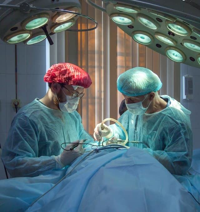
Empyema is usually treatable
Recovery is possible with prompt antibiotics, effective drainage and timely escalation when the infected collection does not resolve.
Antibiotics alone may not be enough
Once pus, loculation or biochemical evidence of complicated pleural infection is present, the infected fluid often needs to be drained.
What is empyema?
The lungs and chest wall are covered by smooth membranes called pleura. A small amount of lubricating fluid normally allows these layers to move easily during breathing.
During pneumonia or another infection, inflammatory fluid may collect in this space. This is called a parapneumonic effusion. Some effusions remain uncomplicated and resolve with antibiotics. Others become infected, develop internal partitions or turn into pus, resulting in complicated pleural infection or empyema.
Pleural effusion
A general term for excess fluid collecting between the lung and chest wall.
Complicated parapneumonic effusion
Infected or highly inflammatory pleural fluid that is unlikely to resolve with antibiotics alone.
Empyema
Frank pus in the pleural space or an established infected pleural collection.
Empyema is different from a lung abscess
Empyema lies outside the lung in the pleural space. A lung abscess is a pus-filled cavity within the lung tissue itself. Both may occur together, but their drainage and treatment pathways differ.
How does a pleural infection develop?
Pleural infection usually progresses through overlapping stages. The speed and severity vary according to the organism, immune response, timing of treatment and underlying health.
Pneumonia inflames the pleura
Infection within the lung irritates the adjacent pleural membranes and causes fluid to collect.
Bacteria enter the fluid
Organisms and inflammatory cells enter the pleural space, lowering glucose and pH while increasing LDH.
Internal partitions form
Fibrin creates septations and locules that divide the fluid into separate pockets and make drainage more difficult.
Organisation may restrict the lung
A thick inflammatory peel can form around the lung, preventing full expansion and sometimes requiring surgical decortication.
Early reassessment is important
Pleural infection can progress despite appropriate initial antibiotics. Persistent fever, inflammation, pain, breathlessness or an undrained collection should prompt review of imaging, drain function and the need for further intervention.
What causes empyema?
Community-acquired pneumonia
Bacterial pneumonia can spread across the lung surface into the adjacent pleural space.
Hospital-acquired infection
Empyema developing during hospital care may involve different, and sometimes more resistant, organisms.
Chest surgery or procedures
Pleural infection may rarely follow thoracic surgery, oesophageal procedures or pleural intervention.
Lung abscess
An abscess close to the pleura can rupture or spread infection into the pleural cavity.
Tuberculosis
TB can occasionally cause chronic pleural infection or tuberculous empyema.
Oesophageal rupture
Leakage from the oesophagus can produce a severe mixed pleural infection requiring urgent specialist treatment.
Bacteria associated with pleural infection
| Clinical setting | Organisms that may be encountered | Important consideration |
|---|---|---|
| Community-acquired pleural infection | Streptococcus anginosus group, other oral streptococci, Streptococcus pneumoniae, Staphylococcus aureus and anaerobic oral organisms | Mixed aerobic and anaerobic infection may occur even when culture identifies only one organism |
| Hospital-acquired pleural infection | Staphylococcus aureus, including MRSA in relevant settings, Gram-negative organisms and anaerobes | Local resistance patterns and recent antibiotic exposure influence empirical treatment |
| Aspiration-associated infection | Oral anaerobes and mixed oral flora | Dental disease, impaired swallowing and reduced consciousness may be relevant |
| Unusual or chronic infection | Mycobacterium tuberculosis, fungi and other uncommon organisms | Travel, immune status and the duration of symptoms guide targeted testing |
A negative culture does not exclude pleural infection
Antibiotics given before sampling and the difficulty of growing anaerobic organisms can reduce culture sensitivity. Pleural-fluid appearance, pH, glucose, LDH, imaging and the overall clinical picture remain important.
Who is more likely to develop empyema?
Older age or frailty
Older or frail patients may have reduced physiological reserve and less typical symptoms.
Diabetes
Diabetes can impair immune responses and increase the severity of bacterial infection.
Alcohol dependence
Alcohol can increase aspiration risk and may coexist with malnutrition or delayed presentation.
Poor dental health
Oral bacteria and anaerobes can contribute, especially when aspiration occurs.
Swallowing problems
Neurological illness, frailty and impaired swallowing can allow infected material to enter the lungs.
Immune suppression
Cancer treatment, transplantation, corticosteroids, HIV and other immune disorders can increase infection risk.
Chronic lung disease
Bronchiectasis, COPD and recurrent aspiration may increase the risk of severe respiratory infection.
Recent hospital care
Surgery, intensive care, ventilation and recent antibiotics can alter the likely organisms.
Delayed pneumonia treatment
A persistent or inadequately controlled pneumonia can progress into complicated pleural infection.
Symptoms and warning signs of empyema
Symptoms commonly overlap with pneumonia. Concern increases when a patient remains unwell or deteriorates despite appropriate antibiotic treatment.
Persistent fever
Fever, chills or sweats may continue despite initial treatment for pneumonia.
Breathlessness
Pleural fluid compresses the lung and can make breathing more difficult.
Pleuritic chest pain
Sharp pain may worsen with deep breathing, coughing or movement.
Persistent cough
Cough may be dry or productive and usually reflects the associated pneumonia rather than pus draining directly from the pleural space.
Fatigue and weakness
Systemic infection and increased work of breathing can cause profound tiredness.
Reduced appetite and weight loss
Prolonged infection may reduce food intake and increase metabolic demand.
Night sweats
Sweating can occur as fever fluctuates, particularly in prolonged or partially treated infection.
Rapid breathing or heartbeat
Tachypnoea and tachycardia may reflect pain, fever, low oxygen levels or sepsis.
Confusion
Older or severely unwell patients may develop confusion, drowsiness or reduced alertness.
When to seek emergency medical help
Call 999 for severe breathing difficulty, blue or grey lips, collapse, severe confusion, extreme drowsiness, intense chest pain, rapidly worsening illness or other signs of possible sepsis. Empyema usually requires urgent hospital treatment.
How is empyema diagnosed?
Clinical assessment
Symptoms, pneumonia treatment, aspiration risk, surgery, immune status and recent antibiotics are reviewed.
Blood tests
Full blood count, CRP, kidney and liver function, clotting and blood cultures may be required.
Chest X-ray
X-ray can identify a pleural effusion, lung collapse or associated pneumonia.
Thoracic ultrasound
Ultrasound confirms fluid, assesses septations and identifies a safe site for aspiration or drainage.
Chest CT
CT defines locules, pleural thickening, lung abscess, obstruction and other causes of treatment failure.
Pleural aspiration
A sample is obtained with a needle and tested for chemistry, cell count, Gram stain and culture.
Ultrasound guidance improves safety
Pleural aspiration and chest-drain insertion should normally be guided by recent thoracic ultrasound so that the collection can be located and important structures avoided.
What do pleural-fluid tests show?
| Finding | What it suggests | Possible implication |
|---|---|---|
| Frank pus | Established empyema | Drainage is generally required if it can be performed safely |
| Positive Gram stain or culture | Bacterial pleural infection | Supports drainage and allows antibiotic refinement |
| pH of 7.2 or lower | High risk of complicated parapneumonic effusion or pleural infection | Intercostal drainage is generally indicated when an accessible collection is present |
| pH above 7.2 but below 7.4 | Intermediate risk | LDH, glucose, fever, fluid volume, CT enhancement and ultrasound septation help determine whether drainage is needed |
| High LDH | Intense pleural inflammation and cellular breakdown | Supports complicated infection when interpreted with pH and other findings |
| Low glucose | Glucose consumption by bacteria and inflammatory cells | Supports complicated pleural infection |
| Neutrophil-predominant cells | Acute inflammatory response | Common in bacterial parapneumonic effusion |
Pleural-fluid pH must be collected correctly
Exposure to air, local anaesthetic or delays in analysis can distort the result. The sample is generally analysed promptly in a blood-gas analyser using an appropriate syringe.
Treatment of empyema
Intravenous antibiotics
Broad initial treatment usually covers likely aerobic and anaerobic organisms and is refined when microbiology becomes available.
Pleural drainage
A chest tube removes infected fluid, reduces bacterial burden and allows the compressed lung to re-expand.
Pain relief
Effective analgesia supports deep breathing, coughing, mobilisation and drain tolerance.
Oxygen and respiratory support
Oxygen, fluid management or more advanced respiratory support may be required in severe illness.
Intrapleural treatment
Combined enzyme and fibrinolytic therapy may improve drainage from selected loculated collections.
Surgical treatment
VATS or open surgery may be needed when drainage is inadequate or organised pleural disease restricts the lung.
Antibiotic treatment
The empirical antibiotic regimen depends on whether infection was acquired in the community or hospital, local resistance patterns, allergy history, kidney function and risk factors for resistant organisms.
Anaerobic cover is commonly required because oral anaerobes may be involved even when routine cultures are negative. Treatment is usually prolonged and often begins intravenously before changing to oral medication once the patient is improving.
Antibiotic duration is individualised
The course depends on drainage, microbiology, inflammatory-marker improvement, fever, imaging and the patient’s overall recovery. A fixed duration is not appropriate for every patient.
Chest-drain treatment
A chest drain is a flexible tube inserted through the chest wall into the pleural collection. It is connected to a drainage system that allows infected fluid to leave the chest.
Image-guided placement
Ultrasound or CT identifies the best accessible pocket and reduces the risk of incorrect positioning.
Small-bore drainage
A small-bore intercostal catheter is commonly used as initial drainage for pleural infection.
Drain flushing
Saline flushing may be used according to local protocol to reduce blockage by thick fluid.
Output monitoring
Drain volume, appearance, patency and any air leak are regularly reviewed.
Repeat imaging
Ultrasound, X-ray or CT may assess whether the collection is resolving and whether the tube is well positioned.
Daily clinical review
Fever, pain, oxygen requirement, blood tests and overall progress determine whether escalation is needed.
A drain can be present but still fail to drain the infection
Thick pus, blocked tubing, poor positioning or separate locules can leave infected fluid behind. Persistent illness requires prompt reassessment rather than assuming that tube placement alone has achieved source control.
Intrapleural treatment with tPA and DNase
Tissue plasminogen activator helps break down fibrinous septations, while DNase reduces the viscosity of infected fluid by breaking down extracellular DNA. Used together, they can improve drainage from loculated pleural infection.
When it may be considered
The chest drain has stopped draining adequately while imaging shows a residual infected collection.
Potential benefit
Improved fluid drainage may reduce the need for surgery in some patients.
Bleeding risk
Anticoagulation, platelet disorders and recent bleeding require careful review before treatment.
The two medicines are used together
Current evidence supports the combination rather than using either tPA or DNase alone for routine pleural-infection drainage. Dosing and safety assessment are managed by the specialist team.
When is surgery needed?
Surgery is considered when infection cannot be adequately controlled with antibiotics, drainage and appropriate intrapleural treatment, or when organised pleural tissue prevents the lung from expanding.
Video-assisted thoracoscopic surgery
VATS uses small incisions and a camera to clear infected material, divide adhesions and remove restrictive pleural tissue where needed.
Decortication
A fibrous peel is removed from the lung surface so that the lung can expand more fully.
Open thoracotomy
Open surgery may be required for advanced organised infection, difficult anatomy or when minimally invasive treatment is not sufficient.
Respiratory and thoracic-surgical review should be coordinated
The timing of escalation depends on clinical stability, drain response, imaging, operative risk and the stage of pleural organisation. Early multidisciplinary discussion can avoid prolonged ineffective treatment.
Complications of untreated or inadequately drained empyema
Sepsis
Infection can produce systemic inflammation, organ dysfunction and circulatory collapse.
Trapped lung
A thick fibrous peel can prevent the lung from fully expanding.
Pleural fibrosis
Residual pleural thickening may contribute to persistent pain, breathlessness or restrictive physiology.
Respiratory failure
Severe infection, pneumonia and lung compression can impair oxygenation and ventilation.
Bronchopleural fistula
An abnormal connection between an airway and pleural space can cause a persistent air leak and complicate healing.
Empyema necessitans
Rarely, infection extends through the chest wall and forms an external soft-tissue collection.
Lung abscess
Infection within the lung can coexist with the pleural collection and may prolong treatment.
Prolonged deconditioning
Extended hospitalisation can lead to weakness, muscle loss and reduced exercise capacity.
Recurrent infection
Residual collections, obstruction or unresolved aspiration may allow infection to return.
Recovery and follow-up after empyema
Recovery can take several weeks or months, especially after severe pneumonia, sepsis, prolonged drainage or surgery.
Clinical review
Fever, cough, chest pain, breathlessness, appetite and energy levels should steadily improve.
Repeat imaging
Follow-up X-ray or CT may confirm resolution and exclude an underlying obstructing lesion or persistent collection.
Lung-function testing
Persistent breathlessness may justify spirometry, gas-transfer testing or exercise assessment.
Physiotherapy
Breathing exercises, mobilisation and rehabilitation can help restore strength and confidence.
Nutritional recovery
Protein, calorie and dietetic support may be needed after prolonged infection or weight loss.
Underlying-cause review
Aspiration, dental disease, immune suppression, malignancy or airway obstruction may require further treatment.
Persistent breathing limitation can be assessed with specialist lung-function testing .
Can empyema be prevented?
Not every case is preventable, but reducing pneumonia risk and treating respiratory infection promptly can lower the likelihood of progression.
Seek early pneumonia assessment
Persistent fever, breathlessness, chest pain or deterioration should not be left untreated.
Complete prescribed antibiotics
Take antibiotics exactly as directed and seek review if symptoms are not improving.
Pneumococcal vaccination
Vaccination is recommended for eligible age and risk groups according to current NHS guidance.
Influenza and COVID-19 vaccination
Preventing viral infection may reduce secondary pneumonia in vulnerable patients.
Improve oral health
Dental treatment and regular oral hygiene may reduce the burden of aspiration-associated oral bacteria.
Assess swallowing problems
Speech-and-language assessment may reduce recurrent aspiration in people with neurological disease or frailty.
Stop smoking
Smoking damages respiratory defences and increases the risk of pneumonia and other lung disease.
Optimise chronic conditions
Good diabetes control and appropriate management of lung disease can improve infection resilience.
Review recurrent infection
Repeated pneumonia may require investigation for aspiration, bronchiectasis, immune deficiency or airway obstruction.
Vaccination reduces risk but does not prevent every pneumonia
Seek medical assessment when significant respiratory symptoms develop, even when vaccinations are up to date.
Frequently asked questions
Is empyema the same as pneumonia?
No. Pneumonia is infection within the lung. Empyema is an infected collection in the pleural space outside the lung, although it most commonly develops as a complication of pneumonia.
Is empyema contagious?
The pleural collection itself is not passed between people. The infection that caused the pneumonia may occasionally be transmissible, depending on the organism.
Can antibiotics alone cure empyema?
Small uncomplicated parapneumonic effusions may resolve with antibiotics. Established empyema, pus, low pleural-fluid pH or loculated infected fluid usually requires drainage as well.
Is chest-drain insertion painful?
Local anaesthetic and pain relief are used, but discomfort can occur during insertion and while the drain remains in place. Pain should be reported so treatment can be adjusted.
How long does a chest drain stay in?
Duration varies according to drainage, imaging, fever, inflammatory markers and lung expansion. It may be several days or longer in complex infection.
What are tPA and DNase?
They are medicines placed through a chest drain to help break down septations and reduce the thickness of infected fluid, improving drainage in selected patients.
Does everyone with empyema need surgery?
No. Many patients improve with antibiotics, chest drainage and sometimes intrapleural treatment. Surgery is used when infection or lung restriction persists despite these measures.
How long does recovery take?
Recovery may take weeks or months. The timeframe depends on infection severity, underlying health, surgery, nutritional status and the degree of deconditioning.
Can empyema return?
Recurrence is possible if infection is incompletely drained or an underlying cause such as aspiration, obstruction or immune suppression remains.
When should I seek emergency help?
Call 999 for severe breathing difficulty, blue or grey lips, collapse, severe confusion, extreme drowsiness, intense chest pain or rapidly worsening illness.
Conclusion
Empyema is a serious infection of the pleural space that most commonly develops after pneumonia. It should be suspected when fever, breathlessness, chest pain or inflammation persists despite appropriate pneumonia treatment.
Diagnosis combines clinical assessment, blood tests, chest X-ray, thoracic ultrasound, CT where needed and analysis of pleural fluid. Frank pus, positive microbiology, low pleural-fluid pH and loculated collections support the need for drainage.
Effective treatment requires both antibiotics and adequate source control. A small-bore image-guided chest drain is commonly used first. Combined tPA and DNase may improve drainage in selected loculated collections, while VATS or open surgery may be required when medical treatment is inadequate.
Recovery often continues after hospital discharge and may involve follow-up imaging, respiratory assessment, rehabilitation, nutritional support and investigation of the underlying cause.
People with persistent pneumonia symptoms, unexplained pleural fluid or recurrent chest infection can seek specialist assessment through the London Chest Specialist chest infection diagnosis and treatment service .
References and further information
- British Thoracic Society. British Thoracic Society Guideline for pleural disease. View the BTS pleural-disease guideline
- British Thoracic Society. BTS Guideline for pleural disease: summary. View the BTS summary
- Northern Ireland Direct. Empyema. Read patient information about empyema
- Bhatnagar M, Monsch GM, Kette S, et al. Pleural infection in adults: integrating medical and surgical management for optimal outcomes. View the contemporary clinical review
- London Chest Specialist. Chest infection diagnosis and treatment. Explore the chest infection service
- London Chest Specialist. Bronchiectasis diagnosis and treatment. Read about bronchiectasis
- London Chest Specialist. Lung-function testing. Read about lung-function tests
Concerned About Pleural Infection or Persistent Pneumonia?
A respiratory consultation can review symptoms, chest imaging, pleural-fluid results, antibiotic treatment and whether further investigation or specialist follow-up is appropriate.