
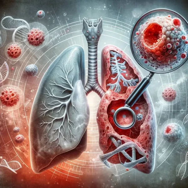
Persistent cough
A cough that does not resolve within three weeks, becomes progressively worse or changes from its usual pattern.
Specialist respiratory assessment for possible lung cancer, suspicious chest symptoms or abnormal imaging, with appropriate investigation and treatment planning based on clinical findings.
Lung cancer develops when abnormal cells in the lungs grow in an uncontrolled way and form a tumour.
Primary lung cancer begins in the lungs. Cancer that has spread to the lungs from another part of the body is known as secondary or metastatic cancer and is managed according to the original cancer type.
The two main categories are non-small-cell lung cancer and small-cell lung cancer. Non-small-cell lung cancer is the more common group and includes adenocarcinoma, squamous-cell carcinoma and other less common subtypes.
The tumour type, molecular characteristics, location, stage and the patient’s overall health all influence which investigations and treatments are appropriate.
Lung cancer is serious because some people do not experience obvious symptoms until the disease is advanced and more difficult to treat.
Many of these symptoms have causes other than cancer. However, a new, persistent, unexplained or worsening symptom should be assessed, particularly when several symptoms occur together.
A cough that does not resolve within three weeks, becomes progressively worse or changes from its usual pattern.
Blood-streaked sputum or larger amounts of blood require prompt clinical assessment.
New or progressively worsening breathlessness during activity or at rest.
Persistent pain, discomfort when breathing or pain associated with coughing.
Persistent or excessive tiredness that is not explained by sleep, activity or another known condition.
Losing weight or appetite without deliberately changing diet or activity.
New wheezing, a persistent voice change or difficulty producing a normal voice.
Swelling of the face, neck or arm, or a drooping eyelid with facial symptoms, requires urgent medical review.
Smoking remains the most important risk factor, but lung cancer can also develop in people who have never smoked.
Tobacco smoke contains substances that damage DNA in lung cells. Risk generally increases with the amount smoked and the length of time a person has smoked, although stopping remains beneficial at any age.
Occupational or environmental asbestos exposure can increase lung cancer risk, particularly when combined with smoking.
Radon is a naturally occurring radioactive gas that can accumulate inside buildings in certain geographical areas.
Repeated exposure to other people’s tobacco smoke can increase risk even when a person does not smoke.
Long-term exposure to air pollution and diesel exhaust may contribute to genetic damage in lung cells.
Having a close relative with lung cancer can increase risk, although inherited mutations account for a minority of cases.
Lung cancer develops when accumulated cellular changes disrupt the normal controls on growth, repair and cell death.
Diagnosis usually requires imaging and, where appropriate, a tumour or lymph-node sample to identify cancer cells under a microscope.
Symptoms, smoking and occupational history, examination and previous imaging are reviewed.
CT identifies the size and position of a lesion and looks for enlarged lymph nodes or possible spread.
The safest biopsy route that can provide diagnosis and staging information is selected.
Further imaging helps assess whether disease is localised or has spread elsewhere.
Specialists review the evidence and agree the diagnosis, clinical stage and recommended treatment options.
CT is more sensitive than a chest X-ray and provides detailed information about a lesion’s size, shape and location.
It can also identify enlarged mediastinal or hilar lymph nodes and abnormalities in the liver, adrenal glands, bones or other areas included in the scan.
A flexible camera is passed through the nose or mouth into the airways. Visible endobronchial abnormalities can be sampled using biopsies, brushings or washings.
EBUS combines bronchoscopy with ultrasound to identify and sample lymph nodes or masses beside the central airways.
It can provide both diagnostic and staging information without the need for an open surgical procedure.
A radiologist uses CT images to guide a needle through the chest wall into a lung lesion and obtain tissue for pathological and molecular analysis.
PET-CT identifies areas with increased metabolic activity and helps determine whether the cancer has involved lymph nodes or distant organs.
Tissue analysis identifies the cancer subtype. Molecular and biomarker testing may determine whether targeted therapy or immunotherapy is likely to be suitable.
In selected circumstances, biopsy may be unsafe, technically impossible or unlikely to change management. The multidisciplinary team can then consider the clinical history, serial imaging, radiological appearance and treatment risk before making a recommendation.
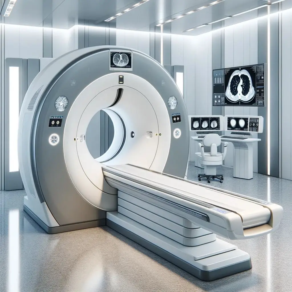
Lung cancer screening uses low-dose CT scanning to look for early cancer in people who are at higher risk but do not have symptoms.
Screening may identify small lung nodules or cancers before they cause symptoms, when potentially curative treatment may be more feasible.
Eligibility depends on factors such as age, smoking history and estimated lung cancer risk. Screening is different from a broader lung health check, which may investigate symptoms, lung function, exercise capacity and general respiratory health.
Produces detailed lung images using a lower radiation dose than a standard diagnostic CT scan.
Smoking history and other personal factors help determine who is most likely to benefit.
Some small nodules require interval CT monitoring rather than an immediate invasive procedure.
Use the lung health check triage tool to compare a general lung health assessment with lung cancer screening and view the available package prices.
A person with coughing up blood, a persistent cough, unexplained weight loss, chest pain or worsening breathlessness should arrange clinical assessment rather than wait for screening.
Once the diagnosis and stage are understood, further tests may be required to determine which treatments can be given safely.
Spirometry, lung volumes and gas-transfer testing assess respiratory reserve before surgery, radiotherapy or systemic treatment.
CPET may be used when surgical risk remains uncertain after standard lung-function testing or when major lung resection is being considered.
Blood count, kidney function, liver function, clotting and other tests can identify treatment risks and provide a baseline before therapy.
An ECG, echocardiogram or cardiology review may be required when there is known heart disease or planned treatment could affect the heart.
MRI or CT of the brain may be required depending on the cancer type, stage, symptoms and proposed treatment.
Mobility, frailty, weight loss, nutrition, social support and existing medical conditions can influence treatment tolerance.
Treatment is determined by the cancer type, molecular profile, disease stage, symptoms and the person’s overall fitness.
Surgery can be considered when disease is confined to an area that can be removed and the patient has sufficient respiratory and general fitness.
Highly focused radiotherapy may treat a small localised tumour when surgery is unsuitable or declined.
Anti-cancer medicines may be given before or after local treatment, alongside radiotherapy, or for advanced disease.
Immunotherapy helps the immune system recognise or attack cancer cells and may be used in selected stages and tumour profiles.
Medicines can target specific molecular changes that drive cancer growth when a suitable alteration is identified.
Small-cell lung cancer commonly requires chemotherapy and radiotherapy because it often spreads early. Surgery is reserved for a very small number of carefully staged, early cases.
Depending on stage, response and current treatment standards, preventative cranial irradiation or scheduled brain imaging may also be discussed by the specialist oncology team.
Diagnosis and treatment planning usually involve several specialists reviewing the same clinical evidence together.
The team considers whether the diagnosis is confirmed, whether further biopsy or imaging is required, the clinical stage, the patient’s fitness and the available treatment options.
Whether pathology and imaging provide sufficient evidence.
Whether disease is localised, regionally advanced or metastatic.
Whether treatment aims to cure, control disease or relieve symptoms.
Surgery, radiotherapy, systemic therapy, surveillance or supportive care.
Supportive care can be provided alongside cancer treatment and is not limited to the final stages of illness.
Treatment of contributing causes, breathing strategies, medication, rehabilitation and oxygen where clinically indicated.
Symptom-directed medicines and selected procedures can help reduce persistent cough, chest pain or airway obstruction.
Dietetic assessment may help when appetite, swallowing, treatment effects or unintended weight loss become difficult.
Clinical nurse specialists, counselling, psychological services and cancer charities can support patients and families.
Dr Ricardo José provides consultant assessment for suspicious respiratory symptoms, lung nodules, abnormal scans and possible lung cancer.
Lung cancer investigation often requires coordination between respiratory medicine, radiology, pathology, thoracic surgery and oncology. Dr José reviews the complete clinical picture and helps identify the safest and most informative next step.
Review of chest X-rays, CT scans, PET-CT findings and previous imaging where available.
Consideration of bronchoscopy, EBUS, CT-guided biopsy or alternative tissue-sampling options.
Lung-function and exercise assessment when treatment suitability requires physiological evaluation.
Access to multidisciplinary cancer pathways at Royal Brompton and The London Clinic where clinically appropriate.
Answers to common questions about symptoms, testing, screening and specialist assessment.
Yes. Smoking is the most important risk factor, but lung cancer can occur in people who have never smoked. Other factors can include radon, asbestos, second-hand smoke, air pollution, family history and acquired cellular mutations.
No. Small or hidden lung cancers may not be visible on a chest X-ray. CT scanning is more sensitive and may be required when symptoms remain concerning or the X-ray is abnormal.
No. Many lung nodules are benign scars, healed infections or other non-cancerous abnormalities. Size, shape, growth, smoking history and other risk factors determine whether monitoring, PET-CT or biopsy is appropriate.
Tissue confirmation is generally sought when it can be obtained safely and will influence treatment. In selected cases, an MDT may make a clinical diagnosis when biopsy is unsafe, not feasible or unlikely to alter management.
Screening uses low-dose CT in eligible people at higher risk who do not have symptoms. Diagnostic CT investigates symptoms, abnormal examinations or suspicious findings and may use a different scan protocol.
The scan is reviewed in the context of the patient’s history and previous imaging. The next step may be repeat CT surveillance, PET-CT, bronchoscopy, EBUS, CT-guided biopsy, blood tests or MDT discussion.
Yes. Depending on cancer type and stage, treatment may include stereotactic radiotherapy, conventional radiotherapy, chemotherapy, immunotherapy, targeted therapy or combinations of these treatments.
Coughing up blood requires prompt medical assessment. Seek emergency help for a significant amount of blood, severe breathlessness, collapse, chest pain or rapidly worsening symptoms.
Arrange a consultant respiratory review for suspicious symptoms, lung nodules, abnormal chest imaging or investigation of possible lung cancer.