
Reflux, aspiration and respiratory disease
Impact of Reflux and Aspiration on Lung Disease: Mechanisms, Patterns of Injury, and Clinical Management
Reflux and aspiration can contribute to cough, wheeze, recurrent infection and longer-term airway injury—even when heartburn is absent. Understanding the mechanism and pattern of lung damage is essential before deciding how it should be treated.
- Microaspiration
- Aspiration pneumonia
- Aspiration pneumonitis
- Chronic cough
- Bronchiectasis
- Interstitial lung disease
Aspiration may be silent
Some patients experience cough, hoarseness, recurrent infection or worsening lung disease without typical heartburn or obvious choking.
Acid suppression does not stop aspiration
Proton pump inhibitors reduce acidity. They do not necessarily prevent reflux events, non-acid reflux or the physical movement of material into the airway.
How reflux and aspiration can affect the lungs
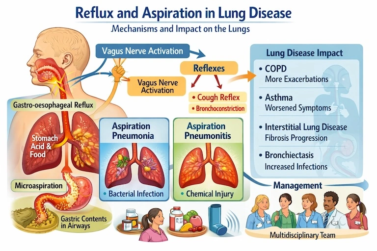
Reflux and aspiration lung disease occur when gastric contents, food, fluid, saliva or oropharyngeal secretions reach the larynx, trachea or lower airways and contribute to respiratory symptoms or tissue injury.
The material reaching the airway may contain gastric acid, pepsin, bile salts, food particles, saliva and bacteria. Its respiratory effects depend on the volume, frequency, chemical composition, bacterial load and the patient’s ability to cough and clear the aspirated material.
A large aspiration event may cause sudden chemical pneumonitis or infection. Repeated small-volume aspiration may be less dramatic but can sustain airway inflammation, impair mucus clearance and worsen an existing respiratory condition.
Importantly, the presence of gastro-oesophageal reflux does not automatically prove that reflux is causing the lung problem. Reflux is common in the general population and in people with chronic respiratory disease. Clinical interpretation therefore requires a plausible pattern, suitable investigations and response to treatment rather than symptoms alone.
Reflux, aspiration and lung disease can reinforce one another. Lung disease may promote reflux, while reflux or aspiration may aggravate respiratory symptoms and airway injury.
The main mechanisms linking reflux to respiratory disease
Direct microaspiration of gastric contents
Small-volume aspiration may occur during sleep, after meals or when lying flat. Repeated exposure to acid, pepsin, bile salts and food material can irritate the airway epithelium and impair mucociliary clearance.
Chemical injury and sterile inflammation
Acidic or bile-containing refluxate can increase epithelial permeability, impair surfactant, provoke neutrophilic inflammation and increase bronchial reactivity.
Aspiration of bacteria-rich secretions
Aspiration does not always begin in the stomach. Saliva and oropharyngeal secretions may contain substantial bacterial loads, particularly with poor dentition, dry mouth or inadequate oral hygiene.
Oesophago-bronchial reflexes
Reflux in the oesophagus may trigger vagally mediated cough or bronchoconstriction without material entering the lungs. This can contribute to cough, wheeze and nocturnal symptoms.
Mechanical interaction with lung disease
Hyperinflation, altered diaphragmatic mechanics, chronic cough, posture and increased work of breathing can alter pressure gradients and potentially promote reflux.
Why heartburn may be absent
Some people experience predominantly extra-oesophageal symptoms, such as cough, throat clearing, hoarseness or wheeze. Reflux can also occur during sleep, when the patient is unaware of it.
Conversely, respiratory symptoms alone are not proof of reflux. Asthma, rhinitis, inducible laryngeal obstruction, infection and other causes still require consideration.
Aspiration pneumonia, aspiration pneumonitis and chronic microaspiration
Aspiration pneumonia
Aspiration pneumonia is an infection that develops after aspiration of bacteria-containing secretions, often from the mouth or upper airway.
It is more common in people with dysphagia, neurological disease, impaired consciousness, frailty, poor oral health or ineffective cough.
Aspiration pneumonitis
Aspiration pneumonitis is an acute inflammatory lung injury caused by aspiration of gastric contents, often following a larger or witnessed aspiration event.
It may produce sudden breathlessness, low oxygen levels, bronchospasm and new infiltrates. The initial process is chemical rather than bacterial.
Chronic microaspiration
Repeated small aspiration events may contribute to chronic cough, laryngeal irritation, airway inflammation, recurrent exacerbations and progressive structural lung changes.
Because aspiration may be silent, diagnosis often depends on risk factors, imaging patterns and targeted testing rather than one symptom.
| Feature | Aspiration pneumonia | Aspiration pneumonitis | Chronic microaspiration |
|---|---|---|---|
| Main process | Bacterial infection | Chemical inflammatory injury | Repeated low-volume airway exposure |
| Typical onset | Hours to days | Often rapid after an aspiration event | Gradual, recurrent or intermittent |
| Possible features | Fever, purulent sputum, consolidation | Hypoxaemia, wheeze, diffuse or dependent infiltrates | Cough, throat clearing, wheeze, recurrent flares |
| Initial management | Antibiotics plus prevention of further aspiration | Supportive care and clinical reassessment | Identify cause, reduce exposure and manage lung effects |
Why pneumonitis and pneumonia can be difficult to distinguish
Chemical pneumonitis can cause fever, inflammatory markers and radiographic shadowing that resemble infection. Secondary bacterial infection may also develop later.
Treatment therefore depends on the clinical setting, severity, time course and reassessment rather than one isolated finding.
Patterns of respiratory injury associated with chronic aspiration
No single CT pattern proves aspiration. However, particular combinations may increase suspicion when they occur alongside suitable clinical risk factors.
Imaging findings must be interpreted alongside swallowing function, reflux risk, microbiology, symptom timing and alternative diagnoses.
Impact on specific respiratory diseases
COPD
Gastro-oesophageal reflux is a frequent comorbidity in COPD and observational studies consistently associate it with more frequent exacerbations.
OR 5.37 Reported association between reflux and COPD exacerbation risk in one observational meta-analysisThis does not prove reflux causes every exacerbation. It does support evaluating reflux and aspiration in frequent exacerbators whose symptoms remain unexplained despite optimised COPD care.
Asthma
Reflux may contribute through microaspiration, oesophageal reflexes or laryngeal irritation. It may present with nocturnal cough, wheeze or throat symptoms.
Proton pump inhibitors produce limited average respiratory benefit when prescribed to unselected patients. Treatment is more likely to help where reflux symptoms or objective evidence are present.
Interstitial lung disease and IPF
Abnormal reflux has been reported frequently in idiopathic pulmonary fibrosis, often without typical symptoms.
Repetitive epithelial injury from microaspiration is biologically plausible, but causality remains uncertain. Reflux may aggravate fibrosis, while restrictive lung mechanics may also promote reflux.
Reflux treatment should not be presented as a proven substitute for established antifibrotic management.
Bronchiectasis
Reflux prevalence estimates vary widely depending on whether symptoms, questionnaires or physiological testing are used. Silent reflux is also reported.
11–75% Range reported in bronchiectasis studies using oesophageal physiological testingReflux has been associated with greater symptoms, exacerbations, hospitalisation and worse quality of life, but association alone does not establish that reflux caused the bronchiectasis.
In asthma, COPD, bronchiectasis and interstitial lung disease, reflux may be one contributor among several. Inhaler technique, airway inflammation, infection, upper-airway disease, obesity, sleep apnoea and cardiac disease may also require assessment.
Clinical clues that should raise suspicion of aspiration
Silent aspiration remains possible
Absence of choking does not exclude aspiration. Neurological impairment, frailty and reduced laryngeal sensation can allow aspiration without a strong cough response.
Practical assessment and investigation pathway
No single test can answer every reflux and aspiration question. Investigation should be driven by the suspected mechanism and how the result would change treatment.
- Clarify symptoms and timing Review heartburn, regurgitation, cough after meals, nocturnal symptoms, choking, wet voice, hoarseness and recurrent infection.
- Identify aspiration risk Assess neurological disease, frailty, sedating medication, dentition, dry mouth, swallowing fatigue, posture and level of consciousness.
- Assess the lung disease Review CT distribution, sputum microbiology, lung function, oxygenation and exacerbation pattern.
- Arrange swallow assessment when indicated Speech and language therapy assessment may be followed by videofluoroscopy or fibreoptic endoscopic evaluation of swallowing.
- Use oesophageal testing selectively Twenty-four-hour pH impedance can detect acid and non-acid reflux. Manometry assesses oesophageal motility and supports planning before some interventions.
- Consider bronchoscopy selectively Bronchoscopy may help investigate persistent infection, obstruction, recurrent focal change or unexplained airway abnormalities.
- Review the whole pattern Interpret results collectively rather than diagnosing aspiration from one symptom, scan feature or reflux test.
| Test | What it assesses | Important limitation |
|---|---|---|
| Videofluoroscopic swallow study | Oral and pharyngeal swallowing, penetration and aspiration | Represents swallowing during a limited test period |
| FEES | Laryngeal function, secretion management and swallowing | Does not assess the oesophageal phase directly |
| 24-hour pH impedance | Acid and non-acid reflux events and proximal reflux | Reflux detection does not by itself prove lung injury |
| Oesophageal manometry | Motility and lower oesophageal sphincter function | Does not directly diagnose pulmonary aspiration |
| CT chest | Distribution of airway, parenchymal and dependent injury | No CT pattern is entirely specific for aspiration |
| Sputum culture | Airway bacteria, fungi and mycobacteria | Organisms do not always identify the route of entry |
Why multidisciplinary care is often needed
Respiratory physician
Assesses the lung phenotype, imaging, microbiology, exacerbations and respiratory consequences of aspiration.
Speech and language therapist
Evaluates swallowing safety, aspiration risk, texture, positioning, pacing and compensatory techniques.
Gastroenterologist
Assesses reflux, oesophageal motility, hiatus hernia and the suitability of medical or procedural treatment.
Respiratory physiotherapist
Optimises airway clearance, breathing technique, cough effectiveness and secretion management.
Dietitian
Balances swallowing safety, nutrition, hydration, weight management and treatment burden.
ENT or neurology team
Assesses laryngeal, upper-airway or neurological causes where these contribute to impaired protection or swallowing.
Prevention and treatment of reflux-related aspiration risk
Management should target the suspected mechanism. Acid suppression alone is not a complete aspiration strategy.
Positioning
Avoid lying flat soon after meals and consider head-of-bed elevation when nocturnal reflux is a concern.
Oral hygiene
Good dental and oral care reduces the bacterial burden that may be aspirated from saliva and secretions.
Swallow optimisation
Use pacing, appropriate posture, supervision and texture modification only when supported by a swallowing assessment.
Medication review
Review sedatives, opioids, anticholinergic drugs and medicines that impair alertness, salivation or swallowing where feasible.
Airway clearance
Bronchiectasis, chronic sputum retention and ineffective cough may require respiratory physiotherapy and a personalised clearance plan.
Nutrition planning
Aspiration prevention must be balanced against dehydration, malnutrition, quality of life and the practicalities of feeding.
Lifestyle and meal-related measures
Measures may include avoiding large late meals, remaining upright after eating, reducing individual dietary triggers, limiting excess alcohol and addressing obesity where relevant.
These measures should be proportionate. Highly restrictive diets can worsen nutrition without improving respiratory outcomes.
Proton pump inhibitors
PPIs are effective for acid-mediated reflux symptoms and oesophagitis. They reduce the acidity of refluxate but do not necessarily reduce the number of reflux events or prevent aspiration.
A time-limited therapeutic trial should have a clear goal and should be reviewed. Continuing treatment indefinitely without benefit may expose the patient to unnecessary medication.
Prokinetic treatment
Prokinetic medicines may be considered in selected patients with impaired motility or gastric emptying, but adverse effects and limited long-term evidence restrict routine use.
Anti-reflux surgery
Procedures such as fundoplication may be appropriate for selected patients with objectively demonstrated reflux and a suitable physiological and surgical profile.
Surgery should not be offered solely because respiratory symptoms coexist with reflux. Multidisciplinary selection and realistic treatment goals are essential.
The respiratory physician’s role in reflux and aspiration lung disease
Patients often present to respiratory services because their main symptoms are cough, wheeze, sputum, breathlessness or recurrent infection rather than heartburn.
The respiratory physician’s role commonly includes:
When urgent medical assessment is needed
Seek urgent help for severe or rapidly worsening respiratory symptoms
Call 999 or seek emergency assessment for severe breathlessness, blue or grey lips, confusion, collapse, severe chest pain, inability to speak because of breathlessness or a major witnessed aspiration followed by respiratory distress.
- Rapidly falling oxygen levels
- New severe wheeze or bronchospasm
- High fever with breathlessness or confusion
- Repeated choking with inability to protect the airway
- Suspected aspiration in a patient with reduced consciousness
Conclusion
Reflux and aspiration can affect the lungs through direct microaspiration, chemical injury, bacterial seeding, oesophago-bronchial reflexes and interactions with existing lung disease.
The clinical consequences range from acute aspiration pneumonitis and aspiration pneumonia to chronic cough, airway inflammation, recurrent exacerbations and structural lung damage.
Reflux is common, and its presence does not automatically prove causation. The strongest clinical approach combines pattern recognition, swallowing assessment, suitable reflux testing, respiratory investigations and multidisciplinary review.
Treatment should address aspiration risk, oral hygiene, swallowing, positioning, nutrition and airway clearance—not simply prescribe acid suppression and assume the problem is resolved.
Frequently asked questions
Can reflux cause lung scarring?
Repetitive microaspiration may contribute to epithelial injury and fibrosis in selected patients, particularly in discussions surrounding idiopathic pulmonary fibrosis. However, causality is not fully established, and treating reflux is not a guaranteed method of slowing fibrotic lung disease.
How can aspiration pneumonitis be distinguished from aspiration pneumonia?
Pneumonitis often follows a witnessed or suspected aspiration event and may cause rapid breathlessness, hypoxaemia and inflammatory changes. Pneumonia is an infection and more often produces persistent fever, purulent sputum and evolving consolidation. The distinction can remain difficult, so clinical reassessment is important.
Does a proton pump inhibitor stop aspiration?
No. A PPI reduces gastric acidity. It does not necessarily stop reflux events, non-acid reflux or the movement of material into the airway.
Why can chest symptoms occur without heartburn?
Reflux may be silent, occur during sleep or present mainly through cough, throat clearing, hoarseness or wheeze. Aspiration from swallowing impairment can also occur without gastric reflux or typical heartburn.
Can reflux worsen asthma?
Reflux may aggravate asthma symptoms through airway exposure or oesophageal reflexes. However, poorly controlled asthma should not automatically be attributed to reflux. Inhaler adherence, inflammation, allergy, obesity and laryngeal disorders may also need assessment.
Can aspiration cause bronchiectasis?
Recurrent aspiration caused by dysphagia or reflux is a recognised cause of bronchiectasis. Because reflux is common, the diagnosis should be supported by clinical risk factors, imaging distribution, swallowing assessment or other relevant evidence.
Which specialists may be involved?
Care may involve a respiratory physician, speech and language therapist, gastroenterologist, respiratory physiotherapist, dietitian, ENT specialist, neurologist and specialist nursing team.
References and further information
- Hill AT, et al. British Thoracic Society guideline for bronchiectasis in adults. Thorax. 2019. View the guideline
- McDonnell MJ, et al. Current therapies for gastro-oesophageal reflux in the setting of chronic lung disease. ERJ Open Research. 2020. View the review
- Morice AH, et al. ERS guidelines on the diagnosis and treatment of chronic cough in adults and children. European Respiratory Journal. 2020. View the guideline
- Althoff MD. Gastroesophageal reflux, atopic dermatitis, and asthma. American Journal of Respiratory and Critical Care Medicine. 2023. View the commentary
- Lee AL, et al. Gastro-oesophageal reflux in non-cystic fibrosis bronchiectasis. Pulmonary Medicine. 2011. View the review