
Bronchiectasis, immunity and recurrent infection
Bronchiectasis in Immunocompromised Individuals
Bronchiectasis can develop or progress when weakened immune defences allow respiratory infections to recur. Effective care requires more than antibiotics alone: clinicians must investigate the immune problem, improve mucus clearance, treat infection and reduce avoidable immunosuppression.
- Antibody deficiency
- Immunoglobulin replacement
- Steroid reduction
- Prophylactic antibiotics
- Airway clearance
- Opportunistic infection
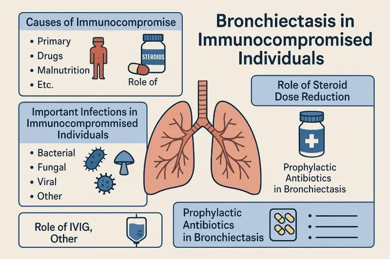
The cause of immune weakness matters
Primary antibody deficiency, blood cancer, HIV, medication and transplantation create different infection risks and require different prevention and monitoring strategies.
Immunosuppression must not be changed abruptly
Steroids and other immune-modifying medicines may be essential. Reductions must be planned with the team treating the underlying condition.
Why immunity is important in bronchiectasis
Bronchiectasis is a permanent widening of parts of the airways. Abnormal airways retain mucus, allowing infection and inflammation to recur. When immune defences are impaired, this infection–inflammation cycle may be more difficult to interrupt.
Repeated or prolonged respiratory infections can damage the airway wall and impair normal mucus clearance. Once bronchiectasis has developed, retained mucus creates an environment in which microorganisms can persist, causing further infection and airway injury.
Immunocompromised patients may experience a wider range of organisms, more severe infection, slower recovery and a greater likelihood of recurrent or opportunistic disease.
Care must therefore address both sides of the problem: the damaged airway and the impaired immune defence.
What does immunocompromised mean?
Being immunocompromised means that one or more parts of the immune system cannot respond to infection as effectively as expected. Immune impairment varies considerably: it may mainly affect antibodies, particular white blood cells, complement, the spleen or several parts of the immune response.
Primary immunodeficiency
Primary immunodeficiencies—also called inborn errors of immunity—arise from an underlying genetic or developmental immune disorder.
- Common variable immunodeficiency
- X-linked agammaglobulinaemia
- Specific antibody deficiency
- Selected IgG-subclass deficiencies
- Combined or cellular immune deficiencies
Secondary immunodeficiency
Secondary immune impairment develops because of another condition, treatment or physiological factor.
- Steroids and other immunosuppressive medicines
- Haematological cancers and their treatments
- Solid-organ or stem-cell transplantation
- HIV infection
- Protein loss, malnutrition or splenic dysfunction
- Age-related changes in immune function
Immunoglobulin concentrations, vaccine responses, lymphocyte populations, medication exposure and infection history must be interpreted together by an appropriately experienced clinician.
Why bronchiectasis develops in immunocompromised people
Infection is not cleared promptly
Bacteria, viruses or fungi may remain in the respiratory tract for longer than they would in a person with intact immunity.
Inflammation persists
Ongoing infection recruits inflammatory cells and enzymes that can injure the airway wall.
Airway structure is damaged
Repeated injury can permanently widen the bronchi and disturb normal ciliary mucus transport.
Mucus accumulates
Widened airways retain sputum, particularly when cough strength or physical activity is reduced.
Microorganisms persist
Retained secretions provide a favourable environment for chronic or recurrent infection.
The cycle repeats
Further infection and inflammation cause additional airway injury and may accelerate disease progression.
This is sometimes described as a vicious vortex: impaired clearance, infection, inflammation and structural damage reinforce one another rather than occurring as a simple one-way sequence.
Infections that may affect immunocompromised patients with bronchiectasis
The likely organism depends on the type and severity of immune impairment, previous antibiotics, hospital exposure, airway structure and geographical or environmental exposures.
Common bacterial pathogens
Haemophilus influenzae, Streptococcus pneumoniae, Moraxella catarrhalis and Staphylococcus aureus may cause exacerbations.
Pseudomonas aeruginosa
Chronic infection is associated with a greater exacerbation burden and may lead to eradication or long-term inhaled antibiotic strategies.
Nontuberculous mycobacteria
NTM pulmonary disease requires species identification, multidrug treatment and specialist microbiological assessment.
Aspergillus and other fungi
Aspergillus can cause sensitisation, ABPA, colonisation, invasive infection or chronic pulmonary aspergillosis.
Respiratory viruses
Influenza, RSV, SARS-CoV-2 and other viruses may provoke exacerbation or severe lower-respiratory disease.
Opportunistic infection
Severe cellular immune impairment may permit infections such as Pneumocystis jirovecii, Nocardia or cytomegalovirus.
A positive sputum result must be interpreted clinically
Detection of an organism does not always prove that it is causing active disease. Symptoms, CT appearances, inflammatory markers, immune status and repeated cultures help distinguish infection, chronic airway infection and incidental detection.
How immune deficiency is investigated in bronchiectasis
All adults diagnosed with bronchiectasis should have a basic assessment for antibody deficiency. More detailed testing is guided by infection history, family history, medication and the wider clinical picture.
Some patients need review by a clinical immunologist or haematologist. A single mildly reduced immunoglobulin result may need repeating and should not automatically lead to immunoglobulin treatment.
Read more about bronchiectasis diagnosis and specialist treatment .
Reducing steroid and immunosuppressive exposure safely
Corticosteroids such as prednisolone suppress inflammation but can also impair immune responses. Infection risk generally rises with higher doses, longer treatment duration, older age, comorbidity and combination immunosuppression.
Some patients with bronchiectasis still need steroids for another condition, including severe asthma, allergic bronchopulmonary aspergillosis, vasculitis, inflammatory disease or transplant medicine.
Steroids must not be stopped abruptly
Long-term systemic steroid treatment can suppress natural adrenal hormone production. Sudden withdrawal may cause adrenal crisis or a dangerous flare of the condition being treated.
- Use the lowest effective dose for the shortest appropriate time.
- Review whether a steroid-sparing treatment is available.
- Confirm whether the steroid is still controlling an active disease process.
- Taper gradually under the direction of the responsible specialist.
- Consider bone, glucose, blood-pressure and infection monitoring.
There is no dose that guarantees freedom from infection risk. Even relatively low doses may be important when combined with another immunosuppressant or used in a patient with additional risk factors.
Inhaled corticosteroids also need a clear indication
Inhaled corticosteroids are not routine treatment for bronchiectasis alone. They remain appropriate when asthma, selected COPD or another recognised inflammatory indication is present.
Immunoglobulin replacement: IVIG and SCIG
Immunoglobulin replacement supplies pooled human IgG antibodies to patients whose immune system cannot produce adequate or functional antibodies.
Intravenous immunoglobulin
IVIG is infused into a vein, often every three to four weeks. It provides a larger intermittent dose and is commonly delivered in a hospital, infusion unit or supervised home programme.
Possible adverse effects include headache, fever, chills, fatigue and infusion reactions. Rare but important risks include thrombosis, haemolysis, kidney injury and aseptic meningitis.
Subcutaneous immunoglobulin
SCIG is given under the skin in smaller, more frequent doses, commonly weekly. Many patients can administer it at home after training.
Local swelling, redness or discomfort can occur. SCIG often produces steadier IgG levels and fewer systemic infusion reactions than IVIG.
NHS England’s 2025 commissioning policy uses an initial immunoglobulin-replacement dose of approximately 0.4–0.6 g/kg each month for qualifying primary antibody deficiencies. Subsequent dosing is individualised according to clinical response, infection burden and IgG measurements.
The goal is not to reach the same laboratory number for every patient. Clinically meaningful outcomes include fewer infections, fewer antibiotic courses, fewer hospital admissions and stabilisation of lung disease.
Patients with established lung damage may require closer monitoring and dose adjustment to control breakthrough infection, but doses should be titrated to clinical response rather than increased automatically.
Not every antibody problem requires immunoglobulin replacement
Some patients with specific antibody deficiency can be protected with vaccination, rapid infection treatment or antibiotic prophylaxis. Immunoglobulin is generally reserved for clearly defined deficiency with a clinically important infection burden despite appropriate alternatives.
Long-term and prophylactic antibiotics
Preventive antibiotics may be considered when recurrent exacerbations continue despite appropriate airway clearance, vaccination, treatment of underlying causes and optimisation of immune management.
Long-term macrolide treatment
Azithromycin or erythromycin may reduce bronchiectasis exacerbations in appropriately selected high-risk patients. Regimens vary; azithromycin 250 mg three times weekly is one pragmatic starting regimen used in specialist practice.
- Exclude active NTM pulmonary disease before treatment.
- Review ECG and QT-prolonging medicines where indicated.
- Discuss hearing, balance and gastrointestinal effects.
- Monitor liver function and microbiology as appropriate.
Long-term inhaled antibiotics
Inhaled antibiotics may be used for selected patients with chronic Pseudomonas aeruginosa infection and continued exacerbation risk.
Options may include colistimethate, gentamicin, tobramycin or another locally approved treatment. Choice depends on microbiology, licensing, previous response and tolerability.
Immunodeficiency-specific prophylaxis
Some patients require prevention directed at a specific opportunistic infection rather than ordinary bronchiectasis exacerbations.
Co-trimoxazole may be used to prevent Pneumocystis jirovecii pneumonia in defined high-risk settings, but eligibility depends on the immune condition, steroid exposure, chemotherapy regimen, transplantation history or HIV immune markers.
Rapid treatment of breakthrough infection
Preventive treatment does not eliminate infection risk. Patients should have a clear plan for sputum sampling, contacting the clinical team and starting appropriate treatment when symptoms worsen.
| Strategy | Possible indication | Important checks | Review |
|---|---|---|---|
| Long-term macrolide | Continued high exacerbation risk despite standard care | NTM cultures, ECG/QT risk, hearing, liver function and interactions | Usually reviewed at least every six months |
| Inhaled antibiotic | Chronic Pseudomonas infection with continued exacerbations | Bronchospasm, delivery technique, culture and susceptibility | Assess exacerbations, tolerance and continuing need |
| Antibiotic prophylaxis for antibody deficiency | Recurrent bacterial infection despite vaccination and ordinary treatment | Organism pattern, allergies, interactions and resistance | Joint immunology and respiratory review |
| Pneumocystis prophylaxis | Defined severe cellular immune suppression or treatment regimen | Blood count, kidney function, potassium and medicine interactions | Continue only while the relevant immune risk persists |
Preventive antibiotics require specialist oversight
Long-term antibiotics can cause adverse effects, alter the airway microbiome and select resistant organisms. Bronchiectasis guidance recommends specialist initiation and regular review of efficacy, toxicity and continuing need.
Airway clearance remains essential
Correcting immune deficiency or giving preventive antibiotics does not remove mucus that is already retained in widened airways. Airway-clearance treatment remains a core part of care.
Read more about airway-clearance devices and questions to ask your physiotherapist .
A comprehensive management plan
- Confirm bronchiectasis and its extent Use an appropriate thin-section chest CT, clinical history and lung-function assessment.
- Define the immune problem Review immunoglobulins, antibody function, blood counts, medication and the underlying disease.
- Characterise respiratory infection Obtain routine and mycobacterial sputum cultures and assess for fungal or opportunistic disease when indicated.
- Optimise immunity safely Replace immunoglobulin when indicated and minimise avoidable immunosuppression without destabilising the underlying condition.
- Improve mucus clearance Establish an effective physiotherapy, exercise and mucoactive treatment plan.
- Prevent future infection Review vaccination, preventive antibiotics and organism-specific prophylaxis.
- Treat exacerbations promptly Use sputum-guided antibiotics and reassess rapidly if the patient deteriorates or fails to improve.
- Monitor meaningful outcomes Track exacerbations, admissions, sputum organisms, lung function, nutrition and quality of life.
Vaccination, nutrition and infection prevention
Vaccination
Influenza, pneumococcal, COVID-19, RSV and other vaccinations should be reviewed according to age, diagnosis, immune status and national guidance.
Timing around treatment
Vaccine effectiveness and suitability may be affected by immunosuppressive treatment, transplantation or immunoglobulin. Live vaccines may be contraindicated.
Nutrition
Weight loss, protein deficiency and micronutrient deficiency can impair immunity, muscle strength and cough effectiveness.
Oral and dental health
Good oral hygiene may reduce aspiration of pathogenic bacteria and should form part of general infection prevention.
Exposure reduction
Smoking cessation, ventilation and sensible infection-control precautions can reduce airway irritation and exposure.
Early clinical contact
Immunocompromised patients may need assessment earlier during an infection because fever and inflammatory responses can be muted.
Regular monitoring and multidisciplinary care
Follow-up should reflect disease severity, immune diagnosis, infection pattern and treatment. Patients with unstable disease may need substantially more frequent review.
The care team may include a respiratory physician, clinical immunologist, haematologist, infection specialist, respiratory physiotherapist, pharmacist, dietitian and the clinician managing the underlying immune condition.
When urgent medical assessment is needed
Immunocompromised patients can deteriorate quickly
Seek urgent assessment for:
- Severe or rapidly worsening breathlessness
- New confusion, collapse or reduced consciousness
- Blue or grey lips
- Significant coughing of fresh blood
- Chest pain with breathlessness
- Low oxygen levels or a marked fall from baseline
- Persistent fever, rigors or severe weakness
- New respiratory symptoms during chemotherapy, transplantation or intensive immunosuppression
Call 999 for severe breathing difficulty, collapse, heavy bleeding or another immediately life-threatening symptom.
Conclusion
Bronchiectasis in an immunocompromised patient requires careful assessment of both the structural lung disease and the underlying immune problem.
Primary antibody deficiencies, blood cancers, HIV, transplantation and immune-suppressing medicines create different patterns of infection and should not be managed as though they were interchangeable.
Immunoglobulin replacement can substantially reduce bacterial infections when a clinically important antibody deficiency is present. The dose should be individualised according to infection burden, clinical response and laboratory monitoring.
Steroid and immunosuppressant exposure should be reduced only when clinically safe and under supervision. Abrupt withdrawal can be dangerous.
Long-term macrolides, inhaled antibiotics and immune-condition-specific prophylaxis may prevent infection in selected patients, but require specialist initiation and regular review.
Airway clearance, exercise, vaccination, nutrition and prompt treatment of breakthrough infection remain fundamental. With coordinated respiratory and immune care, many patients can reduce exacerbations, preserve lung function and improve quality of life.
Frequently asked questions
Can bronchiectasis be cured?
Established bronchiectasis is a permanent structural change. Treatment cannot return widened airways to normal, but it can reduce infections, improve symptoms and slow further damage.
Does everyone with bronchiectasis need immune testing?
Adults should have serum IgG, IgA and IgM measured as part of the standard assessment. More detailed immune testing depends on the infection pattern and clinical history.
Is immunoglobulin replacement safe long term?
IVIG and SCIG are established long-term treatments for qualifying antibody deficiencies. Most reactions are manageable, but uncommon serious effects can occur, so treatment requires specialist prescribing and monitoring.
Does bronchiectasis always mean that a higher immunoglobulin dose is needed?
No. Bronchiectasis may justify closer monitoring and dose adjustment when infections continue, but treatment is titrated to clinical response and IgG measurements rather than increased automatically.
Do preventive antibiotics cause resistance?
They can select resistant organisms. Specialists balance this risk against the benefit of preventing exacerbations, admissions and further lung injury, with regular microbiology and treatment review.
Why might doctors try to reduce steroid treatment?
Systemic steroids can increase infection, osteoporosis, diabetes, hypertension and other risks. The aim is the lowest dose that safely controls the underlying disease—not an abrupt or unsafe withdrawal.
Does every patient taking steroids need Pneumocystis prophylaxis?
No. The decision depends on steroid dose and duration, the underlying disease, additional immunosuppressants, blood-cell counts and other risk factors. It requires individual specialist assessment.
Can immunoglobulin replacement replace airway clearance?
No. Immunoglobulin improves antibody protection but does not remove mucus retained in widened airways. Both treatments may be necessary.
What can patients do at home?
Follow the prescribed airway-clearance plan, remain active, maintain nutrition, avoid smoking, keep appropriate vaccinations up to date and report changes in sputum, breathlessness or general health promptly.
References and further information
- Chalmers JD, et al. European Respiratory Society clinical practice guideline for the management of adult bronchiectasis. European Respiratory Journal. 2025. View the 2025 ERS guideline
- Hill AT, et al. British Thoracic Society guideline for bronchiectasis in adults. Thorax. 2019;74(Suppl 1):1–69. View the BTS guideline
- NHS England. Clinical Commissioning Policy for the use of therapeutic immunoglobulin in England. 2025. View the NHS England policy
- Herrero-Cortina B, et al. European Respiratory Society statement on airway-clearance techniques in adults with bronchiectasis. European Respiratory Journal. 2023. View the airway-clearance statement